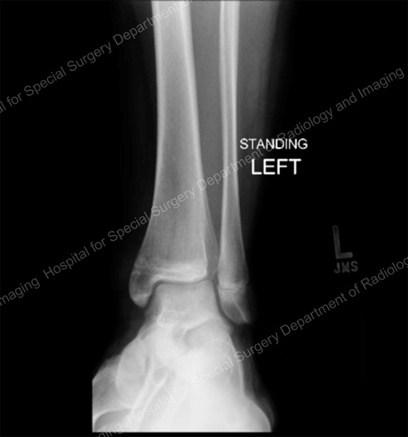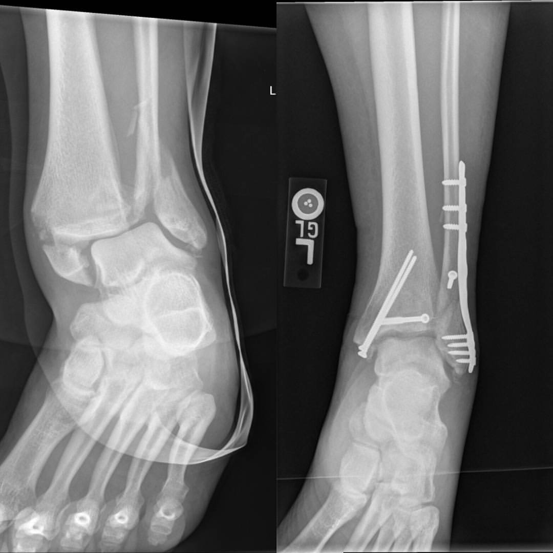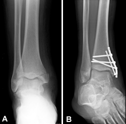

She has mild lateral pain but is non-weight-bearing. Case 3Ī 13-year-old female slips playing soccer. The OAR indicated X-rays, and the emergency department images are above (Case 2, Figure 1). He has mild lateral pain, his medial malleolus is non-tender, and he is non-weight-bearing. Case 2Ī 45-year-old male slips on a sidewalk. The OttawaĪnkle rules (OAR) indicated X-rays, and the emergency department images are above (Case 1, Figure 1). Case 1Ī 22-year-old female falls, injures her ankle, and is non-weight-bearing.

Large or displaced fractures may require operative treatment.Written from the perspective of an emergency physician who also runs a weekly minor fracture clinic, this column is intended to highlight a few key ED teaching points for commonly missed and commonly mismanaged ED orthopedic cases. Small nondisplaced fracture: nonweight-bearing with compressive dressing or NWBSLC for four to six weeks Lateral radiograph (an accessory ossicle, the calcaneus secondarium, may be present) Point tenderness over the calcanealcuboid joint (approximately 1 cm inferior and 3 to 4 cm anterior to the lateral malleolus) Inversion with plantar flexion can lead to an avulsion fracture.įorced dorsiflexion compression fracture. Tenderness to deep palpation between the medial malleolus and the Achilles tendonĭifficult with standard views an oblique ankle radiograph taken with the foot placed in 40 degrees of external rotation has been successful. Posterior talar process (medial tubercle) Large or displaced fragments or persistent symptoms: operative treatment Minimally displaced fracture: NWBSLC for four to six weeks Lateral radiograph (an accessory ossicle, the os trigonum, may be present) Tenderness to deep palpation anterior to the Achilles tendon over posterolateral talus Posterior talar Process (lateral tubercle) Large or displaced fragments: operative treatment Small fragment with <2 mm Displacement: NWBSLC for four to six weeks Mortise view lateral view may show subtalar effusion

Point tenderness over the lateral process (anterior and inferior to the lateral malleolus) Stage I, II, or III (see Table 3): NWBSLC for six weeks Stage IV (see Table 3): surgical treatment Tenderness posterior to the medial malleolus, along the posterior border of the talusĪP view: deep, cup-shaped lesion initial radiograph can be normal because changes in subchondral bone may not develop for weeks. Inversion with plantar flexion or atraumatic Stage III or IV (see Table 3), or persistent symptoms: surgical Treatment Stage I or II (see Table 3): NWBSLC for six weeks Mortise view: shallow, wafer-shaped lesion

Tenderness anterior to the lateral malleolus, along the anterior border of the talus Computed tomographic scans or magnetic resonance imaging may be required because these fractures are difficult to detect on plain films. Delays in treatment can result in long-term disability and surgery. These fractures can often be managed nonsurgically with nonweight-bearing status and a short leg cast worn for approximately four weeks. Posterior talar process fractures are often associated with tenderness to deep palpation anterior to the Achilles tendon over the posterolateral talus, and plantar flexion may exacerbate the pain. Lateral talar process fractures are characterized by point tenderness over the lateral process. Fractures of the talar dome may be medial or lateral, and they are usually the result of inversion injuries, although medial injuries may be atraumatic. However, the clinical presentation of subtle fractures can be similar to that of ankle sprains, and these fractures are frequently missed on initial examination. Most ankle injuries are straightforward ligamentous injuries.


 0 kommentar(er)
